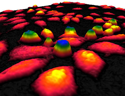
Quantitative Phase Imaging
Usually, to do microscopy (i.e., take an image of a cell), you need some flourescent protein to light up the selected area of the cell(s). However, recent approaches have been made to produce “label-free” images of cells: that is, without the need for some type of (e.g., flourescent) label. One exciting new approach is called quantative phasing imaging (QPI) 1.
The Wikipedia page2 provides a nice synopsis highlighting the advantage over traditional bright-field producing light microscopes:
“Quantitative phase contrast microscopy is primarily used to observed unstained living cells. Measuring the phase delay images of biological cells provides quantitative information about the morphology and the drymass of individual cells. Contrary to conventional phase contrast images, phase shift images of living cells are suitable to be processed by image analysis software. This has led to the development of non-invasive live cell imaging and automated cell culture analysis systems based on quantitative phase contrast microscopy.”3
